Home / Applications / Fluorescence In Vivo Imaging
Camera
Applications
Fluorescence In Vivo Imaging
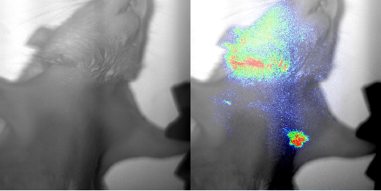
Detection of cancer cells in the lymph nodes of rats
Fluorescence in vivo imaging works on the basis of fluorochromes that are excited by an external light source, and which emit light of a different wavelength in response. The emitted fluorescence can be detected by using a camera which is sensitive in the spectral range of near-infrared. An example is the detection of cancer cells in the lymph nodes of rats. For this purpose, a fluorescence labelled dye is applied intravenously into rats. The detection of the weak emitted fluorescence, which permeates the tissue, requires a highly sensitive camera together with a specific filter.
On the left hand side, a light scatter image of a rat. The picture on the right hand side shows the overlay picture of light scatter image and fluorescence detection of the fluorescence-labelled dye accumulated in the tumour tissue in lymph nodes using the camera GE 1024 1024 DD NIR with a specific filter. The fluorescence around the head of the rat is due to the auto-fluorescence of the fur.
I. Mantouvalou, K. Witte, W. Martyanov, A. Jonas, D. Grötzsch, C. Streeck, H. Löchel, I. Rudolph, A. Erko, H. Stiel and B. Kanngießer, Single shot near edge x-ray absorption fine structure spectroscopy in the laboratory, Appl. Phys. Lett. 108, 201106 (2016)
P. Wachulak, M. Duda, A. Bartnik, A. Sarzyński, Ł. Węgrzyński, M. Nowak, A. Jancarek and H. Fiedorowicz, Compact system for near edge X-ray fine structure (NEXAFS) spectroscopy using a laser-plasma light source, Opt. Express 26, 8260-8274 (2018)